Coinfección de enfermedades zoonóticas transmitidas por garrapatas en humanos del occidente de Venezuela
Introducción. Enfermedades zoonóticas transmitidas por garrapatas, causadas por organismos hemotrópicos de las familias Anaplasmataceae y Babesiidae, se extienden en la mayoría de los países de la region neotropical americana generando graves brotes en la población humana.
Objetivo. En el presente estudio se investigan infecciones y coinfecciones causadas por organismos de los géneros Anaplasma, Babesia y Ehrlichia transmitidas a humanos por picaduras de garrapatas en áreas rurales del occidente de Venezuela donde estas infecciones son endémicas.
Materiales y métodos. Métodos parasitológicos y moleculares (PCR) en muestras de sangre periférica, fueron utilizadas para detectar infecciones por Anaplasma, Babesia y Ehrlichia en poblaciones humanas de áreas rurales del occidente de Venezuela. Un total de 346 muestras sanguíneas fueron evaluadas, incluyendo 226 de pacientes referidos de centros de salud a un centro diagnóstico en Mérida y, 120 de niños escolares y sus representantes muestreados en tres comunidades del estado Barinas.
Resultados. Se registran infecciones y coinfecciones por Anaplasma sp., Babesia sp. y Ehrlichia sp. en los diferentes grupos etarios estudiados. La infección por Anaplasma sp. fue detectada con mayor frecuencia. Anaplasma y Babesia, resultaron infecciones generalizadas y bien adaptadas en el área estudiada y en mayor grado que la causada por Ehrlichia. Particularmente, en el grupo estudiado en el estado Barinas, los análisis estadísticos revelaron una relación epidemiológica en la coinfección Anaplasma-Babesia, evidenciando prevalencias de 92% y 95% con una fuerte asociación en individuos infectados, indicando relación de larga data con las garrapatas vectoras.
Conclusión. Se destaca el significado para la salud pública que tienen las enfermedades transmitidas por garrapatas en el occidente de Venezuela y se sugiere incrementar la vigilancia y control para proteger las poblaciones vulnerables.
ABSTRACT
Introduction. Tick-borne zoonotic diseases, caused by hemotropic organisms of the families Anaplasmataceae and Babesiidae, are of great extend in most countries of the American tropical region generating severe and frequent outbreaks in human populations.
Objective. The present study aimed to investigate the occurrence of tick-borne parasitic infections and coinfections caused by organisms of the genera Anaplasma, Babesia and Ehrlichia in humans exposed to tick-bites through farming or residence in rural areas of Western Venezuela where these infections are endemic.
Materials and methods. Parasitological and molecular (PCR) methodologies in peripheral blood samples were used to detect tick-borne organisms (Anaplasma, Babesia and Ehrlichia) in human populations from rural areas of Western Venezuela. A total of 346 blood samples were evaluated. These included, 226 of patients referred to a diagnostic center in Mérida state coming from different health units and, 120 from schoolchildren and parents sampled in three communities of Barinas state.
Results. The occurrence of infection and coinfection by Anaplasma sp, Babesia sp and/or Ehrlichia sp across different age-groups, was recorded. Anaplasma sp. infection was detected with higher frequency in all the study-groups. Anaplasma and Babesia are widespread and well adapted-infections to a higher degree than Ehrlichia. Particularly, in the group studied from Barinas’ state statistical analyses revealed an epidemiological relationship between Anaplasma and Babesia coinfection, showing the higher prevalence (92% and 95% respectively) and strong association in infected individuals, indicating ancient-relationships with tick-vector.
Conclusion. These findings highlight the significant public health burden of tick-borne diseases in rural Western Venezuela and the need for enhance surveillance and control measures to protect vulnerable populations.
INTRODUCTION
Ticks are the second most common arthropod vectors of human pathogens after mosquitoes, transmitting disease during blood feeding. They can transmit diverse pathogens, including pathogenic bacteria, viruses and parasitic protozoa, several of which cause severe diseases in both humans and animals (1).
Infected animals may exhibit symptoms or remain asymptomatic, while humans typically become accidental hosts through close contact with reservoir animals or in environments where ticks and hosts coexist (2). Notably, approximately 60% of human infections originate from animals, and 75% of emerging infectious diseases are zoonotic (3). Zoonoses pose a significant threat to global health, security, and economic stability, making their prevention a public health priority (4). However, the fundamental mechanisms driving the emergence of zoonoses remain poorly understood (5). Tick-borne diseases caused by bacteria and/or protozoa are prevalent in most countries of the tropical and subtropical regions. Understanding the mechanisms of tick-borne disease transmission under accelerating climate change is imperative (6).
Transmission primarily occurs when ticks feed on host blood (7). Two main pathways exist: systemic transmission and co-feeding transmission. Systemic transmission involves susceptible ticks acquiring infection by feeding on an infected host, or infected ticks transmitting pathogens to a susceptible host during feeding (7,8). In contrast, co-feeding transmission occurs when infected and uninfected ticks feed in close proximity on the same host, enabling direct pathogen transfer between ticks without requiring systemic infection in the host (7,8). Consequently, co-feeding can facilitate pathogen persistence within tick populations even in immune-competent hosts, highlighting that overlooking this pathway significantly underestimates transmission risk (9).
Farming carries an increased risk of zoonotic infections due to close contact with potentially infected animals, particularly as rural populations often depend on livestock for subsistence. Furthermore, as these populations live in close proximity to their livestock, the resulting human-animal interface heightens the risks of exposure and transmission for zoonotic diseases (10,11). Human tick-borne hemoparasitic diseases, including anaplasmosis, babesiosis, and ehrlichiosis, are acute febrile infections caused by pathogens within the genera Anaplasma, Babesia and Ehrlichia. These bacteria and protozoa are major etiologic agents of emerging diseases, increasingly recognized as causes of infection in humans, animals, and tick vectors globally.
Tick-borne hemoparasitic diseases are widely distributed across the Americas, with cases reported from the United States to Argentina. These diseases affect nearly all countries in Central and South America, where various etiologic agents have been detected at high prevalence in livestock, humans, and ticks, frequently during outbreaks (12-14).
At the Center for Parasitological Research, University of Los Andes, Mérida, Western Venezuela, we have frequently received patient referrals over the past two years from hospitals and private clinicians seeking diagnosis for suspected hemoparasitic infections. These symptomatic patients commonly reported abundant ticks and tick bites in their areas of origin, alongside livestock and pet infestations. A similar problem has been documented in rural areas of Barinas, a neighboring state also in Western Venezuela situated in the Llanos region.
The conversion of primary woodlands to grassland ecotones in this region, driven by changing land use patterns, has disrupted natural landscape and biocenoses. This ecological shift, coupled with climate change and a large livestock population, has led to increased tick abundance and the emergence of tick-borne diseases, placing rural communities at persistent risk.
This situation in Mérida and Barinas states prompted the present study to investigate the occurrence of tick-borne parasitic infections caused by Anaplasma, Babesia and Ehrlichia in humans exposed to ticks through farming or residence in rural areas where these infections are endemic in livestock. Our findings revealed a surprisingly high prevalence of affected individuals and frequent detection of both single zoonotic infections and coinfections. This was observed not only among patients referred from health centers but also among children and parents sampled at schools in various localities across Western Venezuela.
PATIENTS AND METHODS
Ethical Approval. This study was approved by the Medical Committee of “Luis Razetti” General Hospital (Barinas, Venezuela; Approval #LRGH-2024-001), the Bioethical Committee of Universidad Francisco de Miranda (Falcón, Venezuela; Approval #UFM-BM-2024-003) and the National Research Council of Venezuela (FONACIT-CFP-2024000182- May 2024). Written informed consent was obtained from all adult participants and legal guardians of minors. Child assent was obtained where age-appropriate. The study adhered to the 2013 Declaration of Helsinki (64th Assembly-Brazil) and Venezuelan biomedical research regulations.
Included Individuals. Between June 2023 and july 2025, 346 participants were enrolled: (i) 226 symptomatic patients showing fever, myalgia, arthralgia and/or headache were referred for hemoparasite diagnostics to the Center for Parasitological Research “J.F. Torrealba”, Universidad de Los Andes (JFT-RC-Mérida) and (ii) 120 asymptomatic individuals from Ticoporo village (T-Barinas), including schoolchildren and parents from “Paolo Freire” (n=47) and “Rómulo Gallegos” (n=49) schools, and volunteers from “Sabana Linda” community (n=24). The inclusion criteria established for individuals enrolled in the protocol from the rural Ticoporo village (Barinas) Western Venezuela were age ≥3-year-old and written consent for all of them. In addition, the excluded criteria were antibiotic use within14 days and immunosuppressive therapy.
Sample Collection: 3 mL peripheral blood was collected via venipuncture into EDTA tubes. Blood extraction was carried out by one of us (Dr. GC) as done during the last 30 years. Blood smears were prepared in triplicate, air-dried, methanol-fixed (3 min), and stained with 10% Giemsa in phosphate buffer (pH 7.2, 40–45 min at 22°C). In addition, two-buffy coat smears were prepared from 80 µL blood sample in 75 mm length capillary tubes centrifugation (3,000xg, 3min) and similarly stained.
Microscopy. Slides were examined at 1,000X magnification under oil immersion, including 500 microscopical fields/patient (300 from blood smears and 200 from buffy coats), using an Axioskop-Zeiss microscope. Intraerythrocytic inclusions (indicative of Babesia sp. and/or Anaplasma sp.) and leukocyte-intracytoplasmic morulae (Ehrlichia sp.) were photographed using Motic 480 camera storing system.
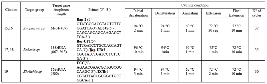
Molecular Analysis. DNA was extracted from EDTA blood using phenol-chlorophorm method after treated with lysis buffer and proteinase K (20 mg/mL). PCR was performed with primers targeting with details provided in Table 1:
Table 1. DNA extraction and details for performed PCR assay
Statistical Analyses. Data were analyzed using: 1. Descriptive statistics (frequencies, percentages) for demographics and infection prevalence. 2. Chi-square or Fisher’s exact tests to compare infection/coinfection rates between groups. 3. Multiple correspondence analysis (MCA) to explore relationships between pathogens (active variables) and demographic/ geographic factors (supplementary variables). Significance was set at p<0.05. Missing data were handled via complete-case analysis.
RESULTS
General Information and Quantification of Tick-Borne Infection
Of the 346 individuals included in this study (226 from JFT-RC-Mérida; 120 from T-Barinas), 307 (88.7%) tested positive for at least one of the detected hemotropic organisms. This included those belonging to Anaplasmataceae (Anaplasma sp. and Ehrlichia sp.) or Babesiidae (Babesia sp.) families. Among the infected individuals, 153 (44.2%) showed Anaplasma-Babesia coinfection; 59 (17.1%) had Anaplasma-Babesia-Ehrlichia coinfection; 15 (4.3%) had Anaplasma-Ehrlichia coinfection; and 10 (2.9%) had Babesia-Ehrlichia coinfection. Additionally, 43 (12.4%) patients had single infections with Anaplasma; 15 (4.3%) with Babesia; and 12 (3.5%) with Ehrlichia.
Considering the total number of infections (including coinfections) for each organism, Anaplasma was detected in 270 individuals (78%), followed by Babesia in 235 individuals (67.9%), and Ehrlichia in 96 individuals (27.7%). The distribution of infections and coinfections in the study population is shown in Figure 1.
Parasitological Observation: Detection and Graphical Record of Hemotropic Organisms
All blood samples (n=346) from referred patients (n=226 /JFT-RC-Mérida) and individuals in endemic areas (n=120/T-Barinas) were microscopically analyzed for hemotropic zoonotic infections. Intra-erythrocytic inclusions indicative of Anaplasma and/or Babesia were detected in Giemsa-stained peripheral blood smears (Figure 2A). Coinfections were frequently observed in the same patients and occasionally within the same erythrocytes (Figure 2B-arrowed). Ehrlichia infection was identified as intracytoplasmic morulae in leukocytes from buffy coat preparations, observed in both single infections (Figure 2A) and coinfections with either Anaplasma or Babesia (Figure 2C). Most infections detected microscopically were confirmed by PCR testing, with representative results shown in Figure 3.
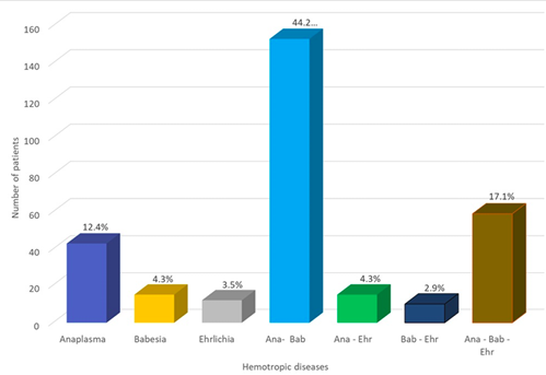
Figure 1. Distribution of hemotropic organisms detected in individuals from different areas of Western Venezuela (note single infection and coinfection among detected organisms).
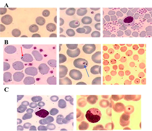
Figure 2. Detection of hemotropic organisms infecting blood cells. A: Single infections with Anaplasma, Babesia, and Ehrlichia. B: Anaplasma-Babesia coinfection. C: Anaplasma-Ehrlichia and Babesia-Ehrlichia coinfections.
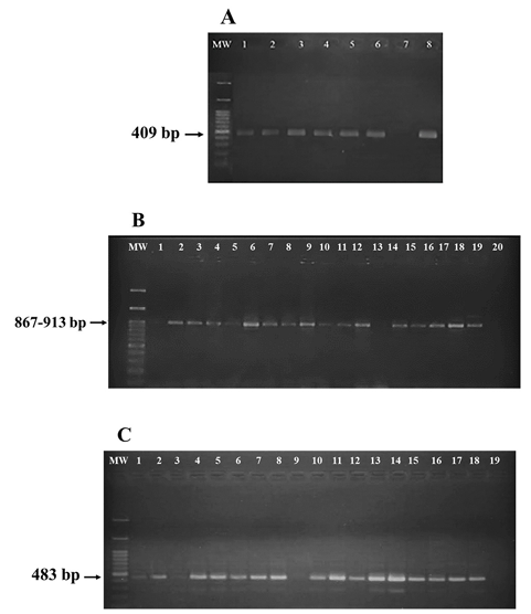
Figure 3. PCR used to corroborate hemotropic infections in patients from different localities of Western Venezuela. A. Anaplasma sp: Lanes 1-6; Lane 7 negative control, Lane 8 positive control. B. Babesia sp. Lanes 1-18; Lane 19 positive control, Lane 20 negative control. C. Ehrlichia sp. Lanes 1-17; Lane 18 positive control, Lane 19 negative control.
Statistical Analyses
Yates’s chi-squared test was used to evaluate independence between pairs of infections. The analysis revealed that Anaplasma infection was not independent of Babesia infection (χ²=55.114, p=1.137×10⁻¹³), demonstrating a statistically significant association between these organisms in infected individuals. This result confirms the frequent Anaplasma-Babesia coinfection observed in the study groups. In contrast, Anaplasma infection was independent of Ehrlichia infection (χ²=0.014, p=0.905), indicating no epidemiological relationship between these pathogens. Similarly, no association was found between Babesia and Ehrlichia infections (χ²=0.503, p=0.479).
Multiple correspondence analysis (MCA) was performed using positive (Ana+, Bab+, Ehr+) and negative (Ana-, Bab-, Ehr-) diagnostic results as active variables. The factorial plane formed by the principal axes (Dim1 and Dim2) represented 80.4% of the total variance. Dim1 was associated with Anaplasma-Babesia coinfection, while Dim2 discriminated the degree of Ehrlichia infection, which appeared as an independent pathology from Anaplasma-Babesia coinfection (Figure 4).
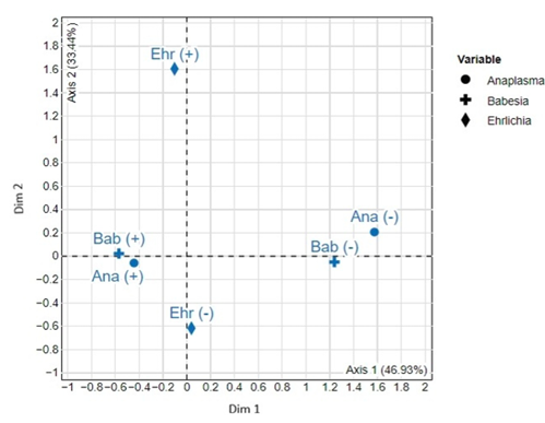
Figure 4. Multiple correspondence analysis of zoonotic hemotropic infections recorded in western Venezuela. Ana (+ve/-ve): Anaplasma; Bab (+ve/-ve): Babesia; Ehr (+ve/-ve): Ehrlichia. Note close Anaplasma-Babesia association in Dim 1.
The proportion of Anaplasma-infected individuals was not independent of gender (χ²=5.069, p=0.024), with higher prevalence in males than females. However, the proportions of Babesia-infected (χ²=1.296, p=0.255) and Ehrlichia-infected (χ²=0.208, p=0.649) individuals were statistically homogeneous.
Age was transformed into an ordinal categorical variable and analyzed using Yates’s chi-squared test. The proportion of Anaplasma-infected individuals was independent of age (χ²=8.200, p=0.414), with homogeneous distribution across age groups. Similarly, age and Ehrlichia infection showed no association (χ²=8.810, p=0.359). In contrast, the proportion of Babesia-infected individuals was not homogeneous across age classes (χ²=23.47, p=0.003)
Prevalence of Zoonotic Hemotropic Infections in the Study Groups
Due to differences in sample collection methods between referred patients with presumptive diagnoses at JFT-Research Center in Mérida state (JFT-RC-Mérida) and blood samples from schools and a community in Barinas’s state (T-Barinas), where tick-borne disease is endemic, prevalence analysis was limited to individuals from the Paolo Freire and Romulo Gallegos schools and the Agua Linda community (T-Barinas). Fisher’s exact test was used instead of chi-squared analysis due to expected cell values below 5. The analysis revealed homogeneous prevalence of Anaplasma (p=0.114), Babesia (p=1.000), and Ehrlichia (p=0.242) across both educational centers and the community, with average values of 92%, 95.8%, and 26.3%, respectively (Table 2). Estimation on the relation of prevalent infection between children and adults, revealed 0.5:1 for Anaplasma and Babesia, and 0.6:1 for infection caused by Ehrlichia sp.
Table 2. Prevalence of zoonotic hemotropic organisms in sampled localities from Barinas’s state, Western Venezuela
|
Sampled locality |
% Prevalence ± SD Infection by Hemotropic Organisms [CI 95% included in each case] |
||
|
Anaplasma |
Babesia |
Ehrlichia |
|
|
Paolo Freire School |
89.4±4.5 [80.7 - 97.7] |
95.7±2.9 [93.1 - 100.0] |
19.1±5.7 [8.0 - 26.3] |
|
Romulo Gallegos School |
98.0±2.0 [94.2 - 100.0] |
95.9±2.8 [90.5 - 100.0] |
34.7±6.8 [21.6 - 45.0] |
|
Sabana Linda community |
87.5±6.8 [75.5 - 98.7] |
95.8±4.1 [88.5 - 100.0] |
25.0±8.8 [9.2 - 34.6]] |
|
Average ± SD |
92± 6.0 [87.3 - 96.6] |
95.8±0.1 [92.3 - 99.2] |
26.3±8.0 [18.7 - 32.7] |
Comparing infections between the 226 referred patients from JFT-RC-Mérida and the 120 individuals from Barinas localities (PF, RG, SL-T-Barinas) revealed: (i) Anaplasma infection was not homogeneous between groups (χ²=21.154, p=4.24×10⁻⁶), with significantly higher prevalence in the T-Barinas group; (ii) Similarly, Babesia infection showed higher prevalence in the T-Barinas group (χ²=61.697, p=4.01×10⁻¹⁵); (iii) Ehrlichia infection was homogeneous between groups (χ²=0.040, p=0.841).
DISCUSSION
This study investigated the frequency and patterns of infection with zoonotic hemotropic organisms (Anaplasma sp., Babesia sp., Ehrlichia sp.) in human populations across Western Venezuela, a region where these pathogens cause known morbidity in livestock and humans (16,18). Our findings reveal an exceptionally high overall prevalence (88.7%) of infection with at least one of these tick-borne pathogens among the 346 individuals studied, comprising both patients referred to a diagnostic center (JFT-RC-Mérida) and residents of an endemic area (T-Barinas). Anaplasma sp. was the most prevalent pathogen (78%), frequently detected as single infections or in coinfection with Babesia sp. (68%) and/or Ehrlichia sp. (28%). Notably, coinfection involving Anaplasma and Babesia was particularly common.
The observed infection rates, especially within the T-Barinas endemic area (Anaplasma: 92±6%; Babesia: 95.8±0.1%), indicate a significant and widespread burden of tick-borne diseases in this region. These rates substantially exceed those typically reported in many other endemic areas (18, 22- 27). The high prevalence of coinfections, particularly the strong statistical association between Anaplasma and Babesia (χ²=55.114, p=1.137x10⁻¹³), suggests a complex epidemiological dynamic. Multiple Correspondence Analysis (MCA) further supported this specific association, implying a potentially long-standing ecological relationship between these two pathogens and their tick vectors in the local environment. In contrast, no significant association was found between Anaplasma/Ehrlichia or Babesia/Ehrlichia coinfections.
Demographic analysis revealed significant variations. Males showed a higher prevalence of Anaplasma infection compared to females (χ²=5.069, p=0.024), while no gender-based differences were observed for Babesia or Ehrlichia. Age-stratified analysis indicated that Babesia infection rates varied significantly across age groups (χ²=23.47, p=0.003), whereas Anaplasma and Ehrlichia prevalence did not show significant age-related differences. Geographically, infection rates for both Anaplasma and Babesia were significantly higher in the T-Barinas endemic area compared to the referred patient group from JFT-RC-Mérida, while Ehrlichia prevalence was similar between both groups (χ²=0.040, p=0.841). The high infection rates (35%-37%) observed among school-aged children in T-Barinas are particularly concerning, given the potential severity of these diseases (22, 23).
Microscopic examination corroborated the molecular findings, frequently revealing high parasitemia with intraerythrocytic inclusions (Anaplasma, Babesia) and leukocyte-intracytoplasmic morulae (Ehrlichia) visible in over 50% of microscopic fields. Double infections (Babesia/Anaplasma within single erythrocytes) were also commonly observed (Fig. 2), providing direct visual evidence consistent with the high PCR positivity rates and coinfection patterns detected.
These results underscore a critical public health challenge in Western Venezuela. The exceptionally high prevalence of Anaplasma, Babesia, and Ehrlichia infections, including frequent coinfections, indicates intense and ongoing transmission cycles involving humans, reservoir hosts, and tick vectors. The clinical presentation of these diseases can be non-specific initially (13,18), leading to delayed diagnosis and treatment, which is particularly dangerous given the potential for severe outcomes, especially in vulnerable populations like children, the elderly, and immunocompromised individuals (22, 23). The strong Anaplasma-Babesia association warrants further investigation into shared vector competence or reservoir dynamics, as well as the specific identification of circulating species in different places of Western Venezuela.
As expressed through the text the infections caused by hemotropic zoonotic organisms have been taxonomically referred as generic. However, the specific species detected will be presented elsewhere in a series of publications including clinical aspects and therapy.
Based on the present findings, we recommend the following actions:
1.Enhanced Surveillance: Implement long-term, integrated surveillance programs across Western Venezuela, combining traditional microscopy with molecular methods (PCR) to monitor pathogen specific-identification and prevalence in ticks, domestic animals, and human populations. This is crucial for understanding transmission dynamics and identifying emerging hotspots both in animals and human.
2.Vector Competence Studies: Conduct definitive studies to confirm the vector competence of locally prevalent tick species for Anaplasma, Babesia, and Ehrlichia pathogens of public health and veterinary importance.
3.Predictive Modeling: Utilize ecological niche modeling, Species Co-Occurrence models and predictive analytics to forecast potential shifts in tick distribution and pathogen transmission risk in response to climate change and land-use modifications (28).
4.Integrated Control Strategies: Develop and implement integrated tick-borne disease control strategies. This should include promoting biological control methods for ticks and conducting targeted health education campaigns for at-risk communities, particularly in rural areas like T-Barinas, to reduce tick bites and improve awareness of early symptoms.
5.Diagnostic and Clinical Awareness: Increase awareness among healthcare providers in the region regarding the high prevalence and potential severity of these tick-borne diseases. Emphasize the importance of considering them in the differential diagnosis of febrile illnesses, even in the absence of a known tick bite history, and the need for prompt diagnosis and appropriate treatment.
In short, this study documents an alarmingly high prevalence of tick-borne Anaplasma, Babesia, and Ehrlichia infections in human populations in Western Venezuela, with significant rates of coinfection, particularly involving Anaplasma and Babesia. The findings highlight an urgent need for coordinated public health and veterinary interventions to mitigate the substantial disease burden identified in these communities.
CONCLUSION
This study demonstrates a high prevalence of tick-borne infections caused by Anaplasma sp., Babesia sp., and Ehrlichia sp. in human populations across Western Venezuela, confirmed by both parasitological and molecular methods. Infection rates were exceptionally high, particularly in the endemic T-Barinas region, with Anaplasma and Babesia being most widespread. A significant epidemiological association was found between Anaplasma and Babesia coinfections, suggesting a complex interplay between these pathogens and local tick vectors. These findings reveal a substantial and under-recognized burden of tick-borne disease in the region, necessitating urgent public health attention and integrated control strategies.
CONFLICT OF INTEREST
The authors declare they have not conflict of interest.
ACKNOWLEDGEMENTS
We thank the collaboration to The Internal Medicine Unit Hospital Universitario de Los Andes- Mérida-Venezuela and The MinCyT (Vice-Ministry and Codecyt) during this study. The financial support received from FONACIT (Project-CFP-2024000182) is grateful acknowledged. We are indebted to Ernesto P.R. Añez-Rojas for support given during redaction of the MS, Dr. G. Bianchi for statistical analyses and Dr. L.F. Chaves, at Indiana University, for carefully reviewing the MS.
REFERENCES
- de la Fuente J, Estrada-Peña E, Venzal JM, Kocan K M, Sonenshine D. Ticks as vectors of pathogens that cause disease in humans and animals. Frontiers in Bioscience; 2008. 13 6938-6946. PMID: 18508706-DOI: 10.2741/3200
- Wilson, M L, G H Adler, and A Spielman. Correlation Between Abundance of Deer and That of the Deer Tick, Ixodes dammini (Acari: Ixodidae). Annals of the Entomological Society of America; 1985. 78: 172-176.). https://doi.org/10.1093/aesa/78.2.172.
- González-Sanz M, Fhogartaigh C N. Zoonotic Infections. Book Chapter; 2019. 35:290-296. Oxford University Press. https://doi.org/10.1093/oso/9780198801740.003.0046.
- Koren, P and Chaves, LF. 2024. https://academic.oup.com/bioscience/advance-article/doi/10.1093/biosci/biaf045/8114226.
- Otte and Pica-Ciamarra. Emerging infectious zoonotic diseases: The neglected role of food animals. One Health;2021. 13:100323. https://doi.org/10.1016/j.onehlt.2021.100323.
- Wang N and Liu S. Modeling of periodic input Ornstein–Uhlenbeck temperature-tick-borne disease transmission coupling mechanism under climate change. Acta Tropica; 2025.261: 107490. https://doi.org/10.1016/j.actatropica.2024.107490.
- Randolph S, Gern, L, Nuttall, P, Co-feeding ticks: epidemiological significance for tick-borne pathogen transmission; Parasitol. Today;1996.12(12):472–479. https://doi.org/1038.1016/S0169-4758(96)10072-7.
- Voordouw MJ. Co-feeding transmission in Lyme disease pathogens. Parasitology; 2015.142 (2), 290–302. http://refhub.elsevie.com(S0001-706x24)00371-1/sb38.
- Nah K, Magpantay F M G, Bede-Fazekas á, Röst G, Trájer A J, Wu X, Zhang X, Wu J. Assessing systemic and non-systemic transmission risk of tick-borne encephalitis virus in Hungary. PLoS One;2019.14(6), e0217206. http://refhub.elsevie.com(S0001-706x24)00371-1/sb20.
- Diuk-Wasser M, Vannier E, Krause PJ. Coinfection by the tick-borne pathogens Babesia microti and Borrelia burgdorferi: ecological, epidemiological and clinical consequences. Trends Parasitol;2016. 32(1): 30–42. doi:10.1016/j.pt.2015.09.008.
- 11. Krause PJ. Human Babesiosis. International Journal for Parasitology.2019.49(2):165-174. https://doi.org/10.1016/j.ijpara.2018.11.007
- Payne RC, Scott JM. Anaplasmosis and babesiosis in El Salvador. Trop. Anim. Health Prod.1982. 14: 75-80. https://www.agronomia.ues.edu.sv/agrociencia.
- Guglielmone A A. Epidemiology of babesiosis and anaplasmosis in South and Central America. Vet Parasitol.1995. Mar;57(1-3):109-19. doi: 10.1016/0304-4017(94)03115-d.
- Rodriguez-Morales AJ, Bonilla-Aldana DK, Idarraga-Bedoya SE et al. Epidemiology of zoonotic tick-borne diseases in Latin America: ¿Are we just seeing the tip of the iceberg? F1000Research; 2019.7:1988 (https://doi.org/10.12688/f1000research.17649.2).
- Stich R, Bantle, JA, Kocan, K M, Fekete, A. Detection of Anaplasma marginale (Rickettsiales: Anaplasmataceae) in hemolymph of Dermacentor andersoni (Acari: Ixodidae) with the Polymerase Chain Reaction. J. Med. Entomol; 1993.30(4):781-788. DOI: 10.1093/jmedent/30.4.781.
- Añez-Rojas N, Romero O, Valbuena H, Crisante G, Rojas A, Bolívar, A M, Añez N. Detection of Anaplasma marginale Transplacental Transmission in Asymptomatic Cattle. Revista Científica, FCV-LUZ; 2010. Vol. XX, Nº 4, 377-382. https://www.redalyc.org/articulo.oa?id=95916179007.
- Alhassan A, Pumidonming W, Okamura M, Hirata H, Battsetseg B, Fujisaki K, Yokoyama N, Igarashi I. Development of a single-round and multiplex PCR method for the simultaneous detection of Babesia caballi and Babesia equi in horse blood. Veterinary Parasitology; 2005.129:4349. PMID: 15817201-DOI: 10.1016/j.vetpar.2004.12.018.
- Añez N, Rojas A, Crisante G, Abello J, Zambrano C, Quiñonez M. Human babesiosis in Western-Venezuela. Case Reports. CientMed; 2020.1(27):1-6. ISSN-e: 2665-0479.
- Murphy G L, Murphy SA, Whitworth LC, Fox JC, Kocan AA. A molecular and serologic survey of Ehrlichia canis, E. chaffeensis, and E. ewingii in dogs and ticks from Oklahoma. Veterinary Parasitology; 1998. 79. 325±339. PMID: 9831955. DOI: 10.1016/s0304-4017(98)00179-4
- Anderson, JF, The natural history of ticks. Med. Clin. 2002. 86 (2): 205–218. http://refhub.elsevie.com(S0001-706x24)00371-1/sb23.
- Chen SM, Dumler JS, Bakken JS, Walker DH. Identification of a granulocytotropic Ehrlichia species as the etiologic agent of human disease. J. Clin. Microbiol.1994.32, 589±595. doi: 10.1128/jcm.32.3.589-595.1994.
- Ismail N, Bloch KC, McBride J W. 2010. Human Ehrlichiosis and Anaplasmosis. 2010; 30: 261–292. doi: 10.1016/j.cll.2009.10.004.
- Krause PJ, Lepore T, Sikand VK, Gadbaw J, Burke G, Telford S, et al. Atovaquone and azithromycin for the treatment of babesiosis. N Eng J Med, 2003; 343(20):1454-1458. doi: 10.1056/NEJM200011163432004.
- Herc E, Pritt B, Huizenga T, Douce R, Hysell M, Newton D, et al., Probable locally acquired Babesia divergens– like infection in woman, Michigan, USA. Emerg Inf Dis, 2018; 24(8):1558-1560. https://doi.org/10.3201/eid2408.180309.
- Homer MJ, Aguilar-Delfín I, Telford S, Krause PJ, Persing DH. Babesiosis. Clin Microbiol Rev; 2000. 13(3): 451–469. https://doi.org/10.1128/cmr.13.3.451
- Melendez R. Babesiosis: An emerging zoonosis in temperate and tropical zones. A Review. Rev. Cient. 2000. FCV-LUZ, 10(1):13-18.
- Gray JS, Weiss LM. (2008) Babesia microti. In: Khan, N. (Ed.), Emerging Protozoan Pathogens. Taylor and Francis, Abingdon, UK, 2008. pp. 303–349. https://doi.org/10.1016/j.ejop.2008.02.001
- Gao Y, Añez N, Chaves LF. High Spatial Resolution Ensemble Species Distribution Modeling of Rhodnius prolixus, Vector of Chagas Disease, in Western Venezuela. GeoHealth, 2026 (In Press).