Macroadenoma hipofisario con presentación como dolor odontogénico y neuralgia del trigémino: retraso diagnóstico
Introducción: Los macroadenomas hipofisarios pueden causar déficits neurológicos progresivos debido a la compresión de estructuras adyacentes. El diagnóstico tardío incrementa el riesgo de complicaciones visuales y endocrinas.
Presentación del caso: Varón de 64 años que consultó por un año de evolución de cefalea unilateral derecha, dolor facial y deterioro progresivo del campo visual. Las evaluaciones iniciales atribuyeron los síntomas a patología odontológica y posteriormente a neuralgia del trigémino. La neuroimagen, realizada más de un año después del inicio de los síntomas, reveló un macroadenoma hipofisario paraselar de 27 × 35 × 31 mm. El paciente fue sometido a resección quirúrgica transcraneal, logrando una resección tumoral subtotal sin déficits neurológicos ni endocrinos posoperatorios. Se administró radioterapia adyuvante debido a tumor residual. El estudio histopatológico confirmó un tumor neuroendocrino hipofisario sin atipia.
Discusión: Este caso ilustra cómo la demora en la realización de estudios de neuroimagen en pacientes con cefalea unilateral persistente y síntomas visuales puede retrasar el diagnóstico de tumores intracraneales, con la consiguiente aparición de morbilidad potencialmente prevenible. La neuroimagen precoz debería considerarse ante la presencia de signos neurológicos de alarma.
ABSTRACT
Introduction: Pituitary macroadenomas may cause progressive neurological deficits due to compression of adjacent structures. Delayed diagnosis increases the risk of visual and endocrine complications.
Case presentation: A 64-year-old male presented with a one-year history of right-sided headache, facial pain, and progressive visual field impairment. Initial evaluations attributed symptoms to dental pathology and later to trigeminal neuralgia. Neuroimaging performed more than one year after symptom onset revealed a 27× 35 × 31 mm parasellar pituitary macroadenoma. The patient underwent transcranial surgical resection, achieving subtotal tumor removal without postoperative neurological or endocrine deficits. Adjuvant radiotherapy was administered due to residual tumor. Histopathological examination confirmed a pituitary neuroendocrine tumor without atypia.
Discussion: This case illustrates how delayed imaging assessment in patients with persistent unilateral headache and visual symptoms may postpone the diagnosis of intracranial tumors, leading to potentially preventable morbidity. Early neuroimaging should be considered when neurological warning signs are present.
INTRODUCTION
Pituitary adenomas are the most common tumors of the hypothalamic–pituitary axis, accounting for approximately 10–15% of intracranial neoplasms in adults (1,2). They arise from clonal proliferation of adenohypophyseal cells driven by hormonal and genetic alterations and are characterized by localized growth without metastatic potential, distinguishing them from pituitary carcinomas (3). These tumors are most frequently located within the sella turcica, a confined osseous structure that predisposes even small lesions to exert compressive effects on surrounding tissues.
According to size, pituitary adenomas are classified as microadenomas (≤10 mm) and macroadenomas (>10 mm) (4,5). Macroadenomas are of particular clinical relevance due to their tendency to extend beyond the sella and compress adjacent structures, including the optic chiasm, cavernous sinuses, and frontal lobes. Such mass effects may result in visual field defects, persistent headaches, cognitive changes, and varying degrees of pituitary dysfunction (6).
Magnetic resonance imaging (MRI) is the diagnostic modality of choice because of its high sensitivity and superior anatomical resolution for assessing tumor size, extension, and relationships with critical neurovascular structures (7). Contemporary evidence further supports its diagnostic value, with MRI demonstrating a sensitivity of approximately 98% for detecting brain tumors (8).
Delayed recognition of pituitary macroadenomas remains a relevant clinical issue, particularly in patients presenting with nonspecific neurological symptoms. Timely and comprehensive evaluation, including early neuroimaging, is essential to reduce the risk of preventable neurological and endocrine morbidity.
CASE REPORT
A 64-year-old male from Puebla, Mexico, with no significant past medical history, presented to the neurology clinic with a one-year history of progressive right-sided headache, facial pain, and visual field impairment. His social history was notable for tobacco use since the age of 20 and alcohol consumption from ages 18 to 40.
Symptoms began in September 2022 following endodontic treatment of a right molar. Shortly thereafter, the patient developed recurrent, prolonged unilateral headaches. Due to the temporal association with dental treatment, he underwent a second root canal procedure without symptomatic improvement. He was subsequently discharged from dental care.
The patient was then evaluated by the neurology service and initially diagnosed with stress-related headache. Despite pharmacological treatment and reduction of daily activities, his symptoms persisted and progressively worsened. Over the following eight months, he was treated with analgesics for headaches with features compatible with tension-type headache. As pain intensity increased and became refractory to nonsteroidal anti-inflammatory drugs, the diagnosis was revised to trigeminal neuralgia, given the unilateral distribution involving the three divisions of the trigeminal nerve. Treatment with tramadol hydrochloride (100 mg/mL oral drops) was initiated.
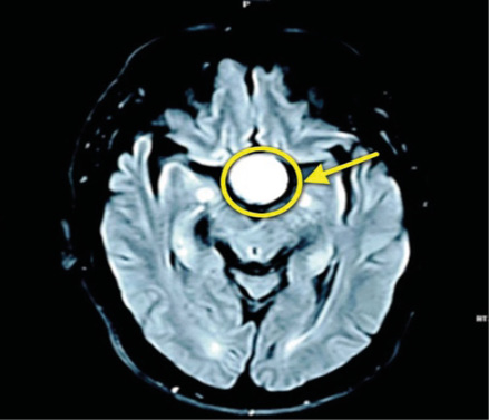
During this period, the patient reported progressive visual impairment, characterized by decreased temporal visual fields bilaterally. This prompted further evaluation, and between September and October 2023, neuroimaging studies were requested. An Axial T2-weighted FLAIR brain MRI revealed a 27× ٣٥ × ٣١ mm parasellar mass consistent with a pituitary macroadenoma (Figure 1).
b)
Figure 1. Axial T2-weighted FLAIR brain MRI demonstrating a well-defined hyperintense lesion in the sellar region with suprasellar extension, exerting mass effect on surrounding structures.
At the time of diagnosis, a neurological examination confirmed visual field compromise. Formal campimetric evaluation demonstrated bitemporal hemianopsia. Baseline pituitary hormonal assessment was performed and showed preserved hormonal function.
After multidisciplinary evaluation, the patient underwent transcranial tumor resection on November 26, 2023, as anatomical characteristics of the sellar floor precluded an endoscopic transsphenoidal approach. The postoperative course was uneventful, with no new neurological or endocrine deficits.
Postoperative CT imaging demonstrated a reduction in both the height and transverse diameter of the lesion. Histopathological analysis of the resected tissue revealed a pituitary neuroendocrine tumor without atypia and no evidence of malignancy (Figure 2).

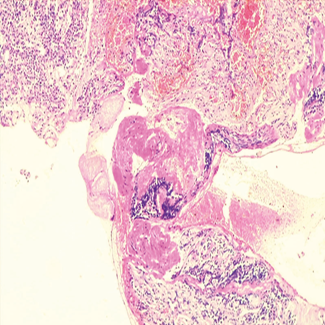
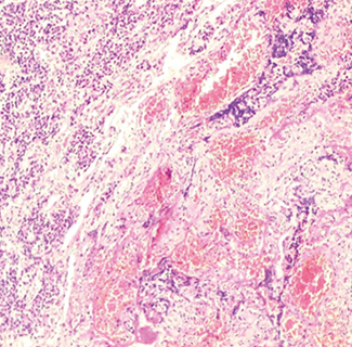
Figure 2. a) Histopathological section stained with hematoxylin and eosin (H&E), low-power magnification (×100). b) Section of a pituitary neuroendocrine tumor measuring 27×35 × 31 millimeters on a slide.
Six months postoperatively, follow-up laboratory studies, including thyroid function tests, were within normal limits. Due to residual tumor, the patient was referred to oncology and received adjuvant radiotherapy consisting of 25 sessions. He continues under annual follow-up with magnetic resonance imaging (MRI), which has shown no evidence of tumor progression. At the most recent evaluation, at age 66, the patient reported marked improvement in symptoms and overall quality of life (Table)
Table. Follow-up laboratory studies
|
Date |
Event |
|
September, 2022 |
First canal root intervention. |
|
November, 2022 |
Diagnosed with odontalgia. Second canal root intervention. |
|
July, 2023 |
Diagnosed with tension-type headaches. |
|
August, 2023 |
Diagnosed with trigeminal neuralgia. |
|
October, 2023 |
Visual field loss (bitemporal hemianopsia). First imaging study is performed. Finding of the tumor. |
|
November, 2023 |
Transcranial surgery. |
DISCUSSION
Despite advances in neuroimaging and increased recognition of pituitary disorders, delayed diagnosis of pituitary macroadenomas continues to be reported, largely due to the nonspecific nature of early symptoms. Headache and subtle visual disturbances are common initial manifestations but are frequently attributed to more prevalent benign conditions, which may delay referral for neuroimaging (9). The absence of early endocrine abnormalities further contributes to diagnostic uncertainty in nonfunctioning tumors.
Headache is widely described as a frequent but nonspecific symptom in patients with pituitary adenomas. Its presence does not consistently correlate with tumor size or degree of invasion, which limits its diagnostic value as an isolated clinical finding. Visual field defects, particularly those resulting from optic chiasm compression, are typically associated with more advanced lesions and often prompt diagnostic imaging. However, gradual visual impairment may go unrecognized by patients, contributing to further delays in diagnosis.
Facial pain and trigeminal nerve involvement are uncommon presentations of pituitary adenomas but have been described in cases with parasellar extension or cavernous sinus involvement. Notably, a review of the available literature reveals a paucity of reports in high-impact journals describing pituitary macroadenomas initially misdiagnosed as dental pathology or primary trigeminal neuralgia. This suggests that such atypical diagnostic pathways are either rare or underreported, despite their potential clinical relevance.
The clinical consequences of delayed diagnosis are primarily related to prolonged compression of surrounding structures. Visual impairment may become partially or completely irreversible when chiasmal compression is sustained over time, even after adequate surgical decompression. Likewise, extended pituitary compression increases the risk of persistent hypopituitarism. Neurological deficits involving cranial nerves may also occur in advanced tumors with extrasellar extension, further impacting patient quality of life.
Although large-scale data quantifying diagnostic delay and associated outcomes are limited, available evidence supports the importance of early imaging in patients with persistent or progressive neurological symptoms.
CONCLUSION.
The functional and secretory characteristics of pituitary tumors, together with the nonspecific nature of early symptoms such as unilateral headache and facial pain, may lead to initial diagnostic interpretations focused on more common neurological or odontogenic conditions. This diagnostic pathway can contribute to delays in definitive evaluation and treatment.
Prolonged tumor growth may result in complications including compression or irreversible damage to the optic pathways, as well as involvement of adjacent bony and neurovascular structures, which can increase surgical complexity and postoperative morbidity. Early recognition of neurological warning signs and timely neuroimaging are therefore essential to facilitate accurate diagnosis, optimize therapeutic planning, and reduce the risk of long-term visual and neurological sequelae.
REFERENCES
- Santaliestra MJC. Tumores hipofisarios. FMC - Formación Médica Continuada En Atención Primaria [Internet]. 1 de enero de 2022;29(1):45-51. Disponible en: https://doi.org/10.1016/j.fmc.2021.05.002
- Castinetti F, Albarel F, Cuny T, Morange I, Vermalle M, Brue T. Adenomas hipofisarios. EMC Tratado de Medicina [Internet]. 2024 Mar 28;28(2):1–11. Disponible en: https://doi.org/10.1016/S1636-5410(24)49086-7
- Jane JA Jr, Catalino MP, Laws ER Jr. Surgical treatment of pituitary adenomas. Endotext [Internet]. 2022. Disponible en: https://www.ncbi.nlm.nih.gov/books/NBK278983/
- Rivera G, Azúa EM, Sánchez JA, Mares CA, Morín A. Macroadenoma hipofisario. Bol Clín Hosp Infant Edo Son. 2020;37(2):123–126.
- Melgar SM, Meléndez M, Díaz E, Barreiro A, Rápalo A, Taboada J, et al. Adenomas hipofisarios al descubierto: el papel imprescindible de la resonancia magnética. Soc Esp Radiol Méd. 2024;1(1).
- Tritos NA, Miller KK. Diagnosis and management of pituitary adenomas: a review. JAMA. 2023;329(16):1386–1398. Disponible en: https://doi.org/10.1001/jama.2023.5444
- Melgar SM, Meléndez M, Díaz E, Barreiro A, Rápalo A, Taboada J, et al. Adenomas hipofisarios al descubierto: el papel imprescindible de la resonancia magnética. Soc Esp Radiol Méd. 2024;1(1).
- Rojas D. Manejo de los tumores de hipófisis. Rev Méd Clín Las Condes. 2017;28(3). Disponible en: https://doi.org/10.1016/j.rmclc.2017.01.008
- Raikwar S, Rao FR. Diagnostic accuracy of CT scan versus MRI in detecting brain tumors. Int J Med Pharm Res. 2024;5(5):344–347. Disponible en: https:/doi.org/10.5281/zenodo.13991350